Importance and Uses of Periodic Acid-Schiff (PAS) Stain
- PAS helps to demonstrate glycogen, cellulose and starch. This is useful to detect glycogen deposits in liver when glycogen storage disease is suspected.
- Basement membrane of various tissues may also be visualized through the PAS stain. The PAS is most commonly used to demonstrate the thickness of glomerular basement membrane when renal disease is being assessed.
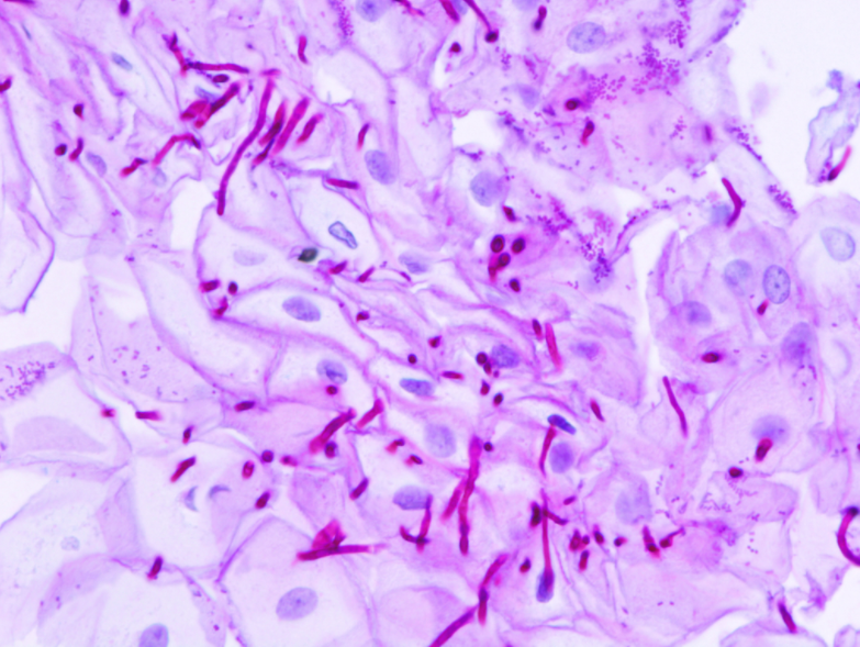
- Certain fungi in tissue samples such as Cryptococcus neoformans, Histoplasma capsulatum, Aspergillus fumiagtus, Blastomyces etc can be demonstrated by PAS stain because of high carbohydrate content in their cell wall/capsule.
- Mucin, particularly acid mucin is demonstrated by PAS. It is important un endocervical glands, intestinal glands and bronchial glands.
- PAS helps to demonstrate cerebrosides and gangliosides. It helps in diagnosis of Gaucher’s disease, Krabbe’s disease and lysosomal storage diseases etc.
- Certain pigments such as lipofuchsin and pigments of Dubin-Johnson syndrome are demonstrated by PAS stain.
- Russell bodies of plasma cells are stained by PAS.
Principle of PAS Stain
Glycol group of carbohydrates are oxidized by periodic acid to release dialdehydes. These dialdehydes on subsequent combination with Schiff’s reagent result in the formation of magenta colored complex, localized at the site of aldehyde formation.
Preparation of Reagents for PAS Stain
Periodic Acid Solution :
Periodic acid solution, used for oxidation is used in any strength between 0.5% and 2.5%. 1% solution is preferred.
Periodic acid= 1gm
Distilled water= 100ml
Schiff’s Reagent
Fuchsin Basic : 1 gm
Distilled water : 100 ml
Sodium metabisulphite : 2 gm
Conc. HCl : 2 ml
Charcoal activated : 0.3 gm
Dissolve basic fuchsin in boiling water, cool at 50°C and filter. Add sodium metabisulphite and HCl. Store at dark room at room temperature overnight. Add charcoal, shake for one minute and filter.
Procedure of PAS Stain
- Deparaffinization: flame the slide on burner and place in the xylene. Repeat the treatment to remove the wax.
- Hydration: Drain xylene and hydrate the tissue section by passing through decreasing concentration of alcohol baths (100%, 90%, 80%, 70%) and water.
- Oxidation: Place the sections in periodic acid solution (1%) for 5-10 minutes.
- Rinse: Wash in at least two changes of distilled water.
- Treatment with Schiff’s reagent: Cover with Schiff’s reagent for 20-30 minutes.
- Rinse: Rinse in running tap water for 5-10 minutes.
- Counterstain: Cover with hematoxylin for 3-5 minutes. Differentiate and blue for the sections.
- Dehydration: Dehydrate in increasing concentration of alcohols.
- Clearing: Put slides in two xylene baths for clearing.
- Mounting: Mount in DPX or other mounting media.
- Observe under microscope.
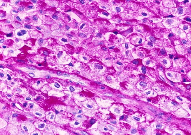
Results and Intrepretation
| Substance | Resulting color |
|---|---|
| PAS positive material | Magenta Pink to Red |
| Nuclei | Blue |
The substances positive for PAS are:
- Polysaccharides: Glycogen, cellulose and starch. Many leucocytes contain glycogen, capsule of fungi (Candida albicans, Histoplasma capsualtum, Cryptococcus and Blastomycosis), actinomycosis and bacteria.
- Glycoproteins: Mucins, mucoid secrection of intestinal tracts, uterine glands, ducts, tracheobronchial tress, hormones (TSH), megakaryocytes etc.
- Glycolipids: Gangliosides, mainly gray matter composed of fatty acids.
- Non-carbohydrate containing substances: Unsaturated lipids, phospholipids and phosphoinositides.
- Certain pigments and substances: Ceroid, lipofucsin, pigment in melanosis coli and Dubin-Johnson pigment.
- Plasmogens: They are acetyl phospholipids, eg: Russell bodies.
- Miscellaneous: Amyloid, cartilage matrix, colloid and ocular lens material.
References
- Shariff, S., & Kaler, A. K. (2016). Principles & Interpretation of Laboratory Practices in Surgical Pathology. JP Medical Ltd.
- Dey, P. (2018). Basic and advanced laboratory techniques in histopathology and cytology. Springer Singapore.
- Bancroft, J. D., & Gamble, M. (Eds.). (2008). Theory and practice of histological techniques. Elsevier health sciences.
- Löffler, H., & Rastetter, J. (2012). Atlas of clinical hematology. Springer Science & Business Media.
- https://www.pathologyoutlines.com/topic/stainspas.html
- https://www.labce.com/spg949466_periodic_acid_schiff_pas_diagnostic_applications.aspx
Be the first to comment