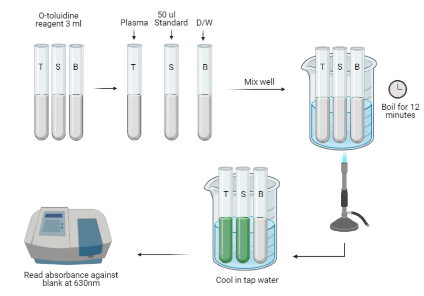
The O-toluidine method is an older method of blood glucose estimation. This method is no longer used today because O-toluidine is believed to be a carcinogen and is replaced by enzymatic methods. This method is still popular because of its simplicity, sensitivity and accuracy.
Principle
The proteins are first precipitated by tricholoroacetic acid. The glucose present in a protein free filtrate react with O-toluidine (primary aromatic amine) in a hot acidic medium to form a stable green colored complex, namely N-glycosamine. The presence of thiourea stabilizes the o-toluidine reaction. The intensity of the color developed is measured photometrically at 630nm, which is directly proportional to the concentration of the glucose present in the fluid.
Requirements
- Apparatus:
Graduated pipettes
Test tubes
Micropipettes
Heating Bath, 100C
Spectrophotometer, wavelength 630nm - Reagents:
Benzoic acid
D-glucose
Glacial acetic acid
O-toluidine
Thiourea
Trichloroacetic acid - Specimen:
Collect 2-3 ml of blood in a fluoride tube. Use plasma for testing. Serum/Whole blood can also be used. Prepare protein-free filtrate if the sample is grossly hemolyzed or icteric. Add 100 ul specimen to 900 ul of 5gm/dl TCA, mix and centrifuge to get the filtrate.
Preparation of Regaents
- Benzoic acid solution 1 g/l:
Dissolve 1 gm of benzoic acid in water and make upto 1 liter. Prepare the benzoic acid at least 24 hours before use. - Stock glucose solution 100 mg/dl:
Dissolve 1 gm of glucose in 1 liter of benzoic acid solution (1 g/l). - O-toluidine reagent:
Dissolve 1.5 gm thiourea in 940 ml of glacial acetic acid. When completely dissolved, add 60ml of O-toluidine. Mix well and store in amber bottle.
Procedure
- Take three (or more if needed) large test-tubes and label as follows:
Blank tube (B)
Standard tube (S)
Test tube (T) - Pipette into each tube as follows:
Test (T) Standard (S) Blank (B) O-toluidine reagent 3 ml 3 ml 3 ml Serum/Plasma 50 μl – – Glucose Standard 100 mg/dl – 50 μl – Distilled water – – 50 μl Note: If protein free filtrate is used instead of serum/plasma, pipette 500 ul of protein free filtrate instead of 50 μl serum/plasma.
- Mix the contents of each tube. Place all the tubes in the water-bath at 100°C for exactly 12 minutes.
- Remove the tubes and allow them to cool in a beaker of cold water for 5 minutes.
- Measure the colour produced in a colorimeter at a wavelength of 630nm.
Calculations
Calculate the concentration of glucose in the blood specimen using the following
formula:

References
- World Health Organization, 2003. Manual of basic techniques for a health laboratory. World Health Organization.
- World Health Organization, 1986. Methods recommended for essential clinical chemical and haematological tests for intermediate hospital laboratories/Working Group on Assessment of Clinical Technologies. In Methods recommended for essential clinical chemical and haematological tests for intermediate hospital laboratories/Working Group on Assessment of Clinical Technologies.
- Mukherjee, K.L., 2013. Medical Laboratory Technology Volume 3 (Vol. 3). Tata McGraw-Hill Education.


Be the first to comment