
Iron deficiency anemia (IDA) may cause a problem in differential diagnosis from other hypochromic anemias like beta-thalassemia trait, alpha-thalassemia trait, HbE disease, sideroblastic anemia or anemia due to chronic diseases. This topic will discuss about laboratory investigations for the differential diagnosis of IDA from those conditions, along with some preliminary investigations.
Hematological Tests
1. Hemoglobin and Hematocrit
According to WHO, the criteria for anemia is when adult males have Hemoglobin levels <13 g/dL and adult females have <12 g/dL. As the iron deficiency worsens, both Hb and PCV decline together.
- Hb >12 g/dl : Not anemic
- Hb 10–11 g/dl : Mild anemia
- Hb 8–9 g/dl : Moderate anemia
- Hb 6–7 g/dl : Marked anemia
- Hb 4–5 g/dl : Severe anemia
- Hb < 4 g/dl : Critical
2. Red Cell Indices
MCV, MCH and MCHC are reduced. RDW is raised.
- Mean Corpuscular Volume (MCV): It is the average volume of the RBC expressed in femtoliters. It becomes <80 fL in IDA (normal 82–98 fL).
- Mean Corpuscular Hemoglobin (MCH): MCH indicates the amount of Hemoglobin (weight) per RBC and is expressed as picograms. MCH will be <25 pg in IDA (normal 27–32 pg).
- Mean Corpuscular Hemoglobin Concentratration (MCHC): The MCHC measures the average concentration of hemoglobin in a red blood cell. MCHC goes below 27 g/dL(normal 31–36 g/dL).
- Red Cell Distribution Width (RDW): RDW is a quantitative measure of anisocytosis. In IDA, RDW is increased and >15%. It is earliest sign of iron deficiency (normal 11.5–14.5%).
Peripheral Blood Smear
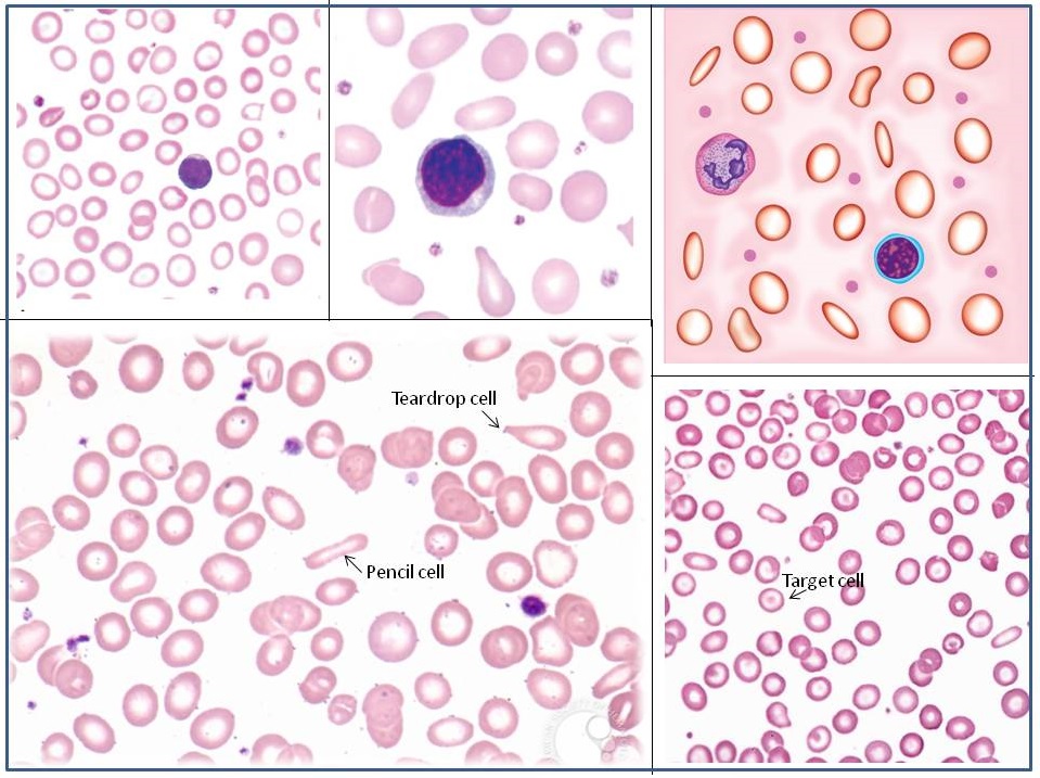
1. Red Blood Cells (RBCs)
- Microcytosis: RBCs are usually smaller than normal. Dimorphic blood picture is seen with a dual population of red cells of which one is macrocytic and the other microcytic and hypochromic when iron deficiency is associated with severe folate or vitamin B12 deficiency.
- Hypochromasia: Central pallor in RBCs is more than 1/3.
- Poikilocytosis: Elliptical forms are common, and elongated pencil (cigar) shaped cells may be seen. Target cells and Teardrop cells may also be present in small numbers. Severe anemia shows ring/pessary cells.
2. White Blood Cells (WBCs)
WBCs are usually normal in number, but can increase due to chronic marrow stimulation in long-standing cases. There may be associated eosinophilia if iron deficiency is secondary to hookworm infestation.
3. Platelets
Platelet count is usually normal, but may be slightly to moderately increased, especially in patients who are bleeding.
4. Reticulocyte Count
The reticulocyte count is low in relation to the degree of anemia.
Bone Marrow Examination
- Cellularity: moderately hypercellular.
- M:E ratio: varies from 2:1 to 1:2 (normal 2:1 to 4:1).
- Erythropoiesis: hyperplasia and micronormoblastic maturation.
- Myelopoiesis: normal.
- Megakaryopoiesis: normal.
- Bone marrow iron: Absent. “Gold standard” test, demonstrated by negative Prussian blue reaction.
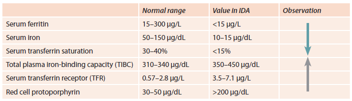
Biochemical Tests
Serum iron profile studies are used to establish a differential diagnosis of microcytic, hypochromic anemia.
Good concept.
HELP ME TO MUCH