
Myeloperoxidase is a lysosomal enzyme present in the primary azurophilic granules of neutrophils, eosinophils and to a certain extent, monocytes. Lymphocytes do not contain myeloperoxidase.
Purpose of Myeloperoxidase stain
Myeloperoxidase (MPO) stain is useful for differentiating the blasts of acute myeloid leukemia (AML) from those of acute lymphoblastic leukemia (ALL). It is also used to diagnose congenital deficiency of neutrophil myeloperoxidase.
Principle of MPO stain
In presence of hydrogen peroxide, myeloperoxidase present in the leukocyte granules oxidize substrates from colorless form to an insoluble blue/brown derivative at the site of the activity. Benzidine or 3,3′-diaminobenzidine or p-phenylenediamine dihydrochloride are used as substrates.
Requirements
- Fixative: 10% formal ethanol or buffered formal acetone.
- Substrate: Benzidine or 3,3′-diaminobenzidine or p-phenylenediamine dihydrochloride
- Buffer: Sorensen’s phosphate buffer, pH 7.3
- 3% Hydrogen Peroxide
- Working substrate: Add 30 mg DAB in 60 ml buffer, add 120 ul hydrogen peroxide and mix.
- Counterstain: Hematoxylin
Procedure of MPO stain
- Fix air dried smears in formal ethanol or buffered formal acetone for 60 seconds.
- Rinse thoroughly in running tap water for 30 seconds.
- Cover the smear with working substrate and incubate for 10 minutes.
- Wash gently with running tap water for 30 seconds.
- Counterstain with hematoxylin for 3-5 minutes.
- Rinse in running tap water and air dry.
- Examine under microscope.
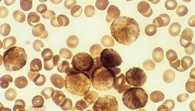
Interpretation
- Myeloperoxidase is present in the primary granules of myeloid cells. Early myeloblasts are negative, with granular positivity appearing progressively as they mature.
- In many cases of AML (without maturation-M1, with maturation-M2 and promyelocytic leukemia-M3), Myeloperoxidase activity has been found in more than 80% blasts. Auer rods are strongly MPO positive.
- Cells of monocytic series display a less intense positive reaxtion that is characterized by fine granular deposits scattered throughout the cell.
- Lymphoblasts and lymphoid cells are MPO negative.
| Positive MPO Reaction | Negative MPO Reaction |
|---|---|
| Neutrophilic granulocytes except blast forms | Basophils (weakly positive) |
| Eosinophils | Lymphocytic cell series |
| Monocytes except blast forms | Erythrocyte cell series |
There is a typo in step 3 of the instructions.
Corrected. Thanks 🙂
Thank you 🙂
Thx
Please,tell me how can I prepare working substrate solution..3,3-diaminobenzadine is not soluble in buffer solution.