Hematoxylin and Eosin (H&E) staining is the most widely used staining technique in histopathology. As its name suggests, H&E stain makes use of a combination of two dyes, namely hematoxylin and eosin. This combination deferentially stains various tissue elements and make them easy for observation.
Principle
The principle behind H & E stain is the chemical attraction between tissue and dye. Hematoxylin, a basic dye imparts blue-purple contrast on basophilic structures, primarily those containing nucleic acid moeties such as chromtatin, ribosomes and cytoplasmic regions rich in RNA. An acidic eosin counterstains the basic elements such as RBCs, cytoplasm, muscle and collagen in varying intensities of pink, orange and red.
Requirements
- Harri’s Hematoxylin stain
A = 1 gm hematoxylin in 10 ml ethanol
B = 20 gm ammonium alum in hot distilled water
Mix A & B, boil and add 0.5 gm of mercuric oxide and filter. - Eosin solution
Yellow eosin = 1 gm
Distilled water = 80 ml
Ethanol = 320 ml
Glacial Acetic Acid = 2 drops - 0.5% HCl
- Dilute ammonia water
Procedure
- Deparaffinization: flame the slide on burner and place in the xylene. Repeat the treatment to remove the wax.
- Hydration: Drain xylene and hydrate the tissue section by passing through decreasing concentration of alcohol baths (100%, 90%, 80%, 70%) and water.
- Nuclear Staining: Stain in hematoxylin for 3-5 minutes.
- Wash in running tap water until sections “blue” for 5 minutes or less.
- Differentiation: selective removal of excess dye from the section). Dip in 1% acid alcohol (1% HCl in 70% alcohol) for a few seconds.
- Blueing: Rinse in running tap water. Dip in ammonia water until the sections become blue, followed by tap water wash.
- Counterstain: Stain in 1% Eosin Y for 10 minutes.
- Wash in tap water for 1-5 minutes.
- Dehydration: Dehydrate in increasing concentration of alcohols.
- Clearing: Put slides in two xylene baths for clearing.
- Mounting: Mount in DPX or other mounting media.
- Observe under compund microscope.
Results and Interpretation
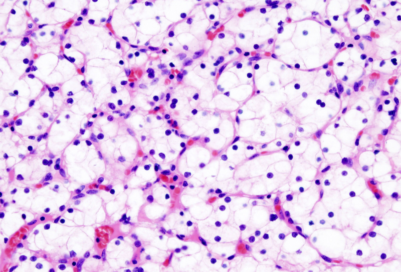
- Nuclei : blue, black
- Cytoplasm : Pink/purplish pink
- Muscle fibres : deep red
- RBCs : orange red
- Calcium : Dark blue
- Mucin : Grey blue
Thanks to meet you.l’m a Microbiologist and want to go into a partner with your Laboratory.Thank you.Keme:AISLT(Nig).
hello that submit very interesting
Can i get the procedure for PAPANICOLOAU STAIN(PAP SMEAR) pls help!