
Sudan Black B (SBB) is a fat soluble dye which has very high affinity for neutral fats and lipids. SBB staining is useful for for the differentiation of Acute myeloid leukemia (AML) from Acute lymphoid leukemia (ALL). It is similar to that of Myeloperoxidase (MPO) staining pattern of leukocytes and monocytes. It has few advantages over MPO:
- SBB can be used for staining smears more than 2 weeks old.
- SBB stains both azurophilic and specific granules in neutrophils, whereas MPO stains azurophilic granules only.
- There is only a little fading of the SBB stain over time.
Principle of Sudan Black B Stain
As SBB is a fat soluble dye, it stains lipids such as sterols, neutral fats and phospholipids. These are present in azurophilic and secondary granules of myelocytic and lysosomal granules of monocytic cells. During staining, the dye leaves the solvent because of its high solubility in lipids than solvent. On microscopic examination, varying degree of black colored pigments are seen in the positive reaction.
Requirements
- Sample: Fresh anticoagulated whole blood or bone marrow smear may be used. The slides must be fixed as soon as possible.
- Fixative: 40% formaldehyde solution vapor
- Stain: SBB 0.3 g in 100 ml absolute ethanol
- Phenol buffer: Dissolve 16 g crystalline phenol in 30 ml absolute ethanol. Add to 100 ml distilled water in which 0.3 g Na2HPO4.12H2O has been dissolved
- Working SBB stain solution: Add 40 ml buffer to 60 ml SBB solution (The composition of working stain may slightly vary upon different products.)
- Counterstain: May–Grunwald–Giemsa or Leishman stain.
Procedure of Sudan Black B Stain
- Fix air dried smears in formalin vapour, formaldehyde or formalin-ethanol fixative for 10 minutes.
- Wash gently in water for 5-10 minutes.
- Place the slides in the working stain solution for 1 hour in a Coplin jar with a lid on.
- Remove and flood the slides with 70% alcohol for 30 seconds. Tip the 70% alcohol off and flood again. Repeat this three times.
- Rinse in running tap water and air dry.
- Counterstain without further fixation with Leishman stain or May–Grunwald–Giemsa stain.
- Air dry and examine microscopically.
Results and Interpretation
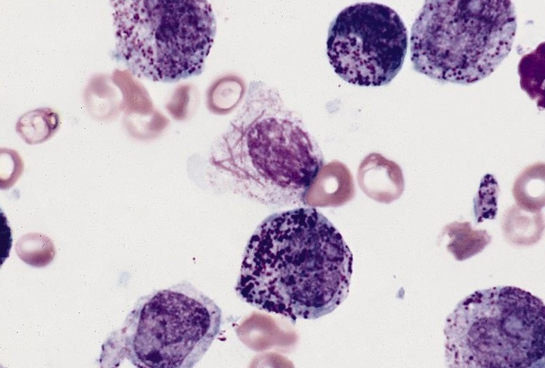
Production of black and granular pigment indicates positive reaction.
- Lipids are present in azurophilic and secondary granules of myelocytic cells. Hence, these are SBB positive. The staining becomes more intense as cells mature from myeloblast to mature forms. Basophils are generally not positive but may show bright red/purple metachromatic staining of the granules.
- Lipids are present in lysosomal granules of monocytic cells. Monocytic cells show variable reactions, from negative to weakly positive.
- Lymphoid cells are SBB negative. However, in ALL, less than 3% of blast cells show positive reaction.
Sudan Black is formed by coupling of diazotized 4-phenylazo-1-naphthylamine with 2,3-dihydro-2,2-dimethyl-1